We are approaching the middle point of our project and want to share a lot of news with you.
In this newsletter we will:
- Show a video describing the overall scope of the project
- Explain what is a Spectral CT, and why it matters to our project
- Give an update on the status of the clinical trials of our project
- Present another two of the partners of the consortium, HCL and UCBL
Video
What is a Spectral CT?
We will start explaining what a CT scan is. CT stands for Computed Tomography and is an imaging technology based on X-rays. X-rays are a type of light with very high energy. Most objects are translucent in this light, that is to say, they allow the light to pass although they might absorb part of it. Bones, for example, absorb more X-ray light than other tissues. For this reason, X-rays are used to take pictures of what is inside our body. If you have ever received an X-ray for a fractured bone, the doctor often will take two images, so that each of the two images show everything inside us from a different perspective. Certain features that may appear in the same spot in one of the images (for example a bone hiding behind a different bone), will be separated in the other one. With both these images, a physician may have a better understanding of what is happening than with a single one. If we increase the number of images we take, such as if we took three images instead of two, we could differentiate even better the spatial distribution of our insides. If we take this approach a step beyond and take an X-ray image from every possible angle in a 360 degree circle around the patient, then we would know perfectly the shape and location of every organ in our patient’s body. With all this information, it is possible to use a Computer (the C in CT) to transform this set of images into a single 3D image. This is the fundamental operation of a CT scan.
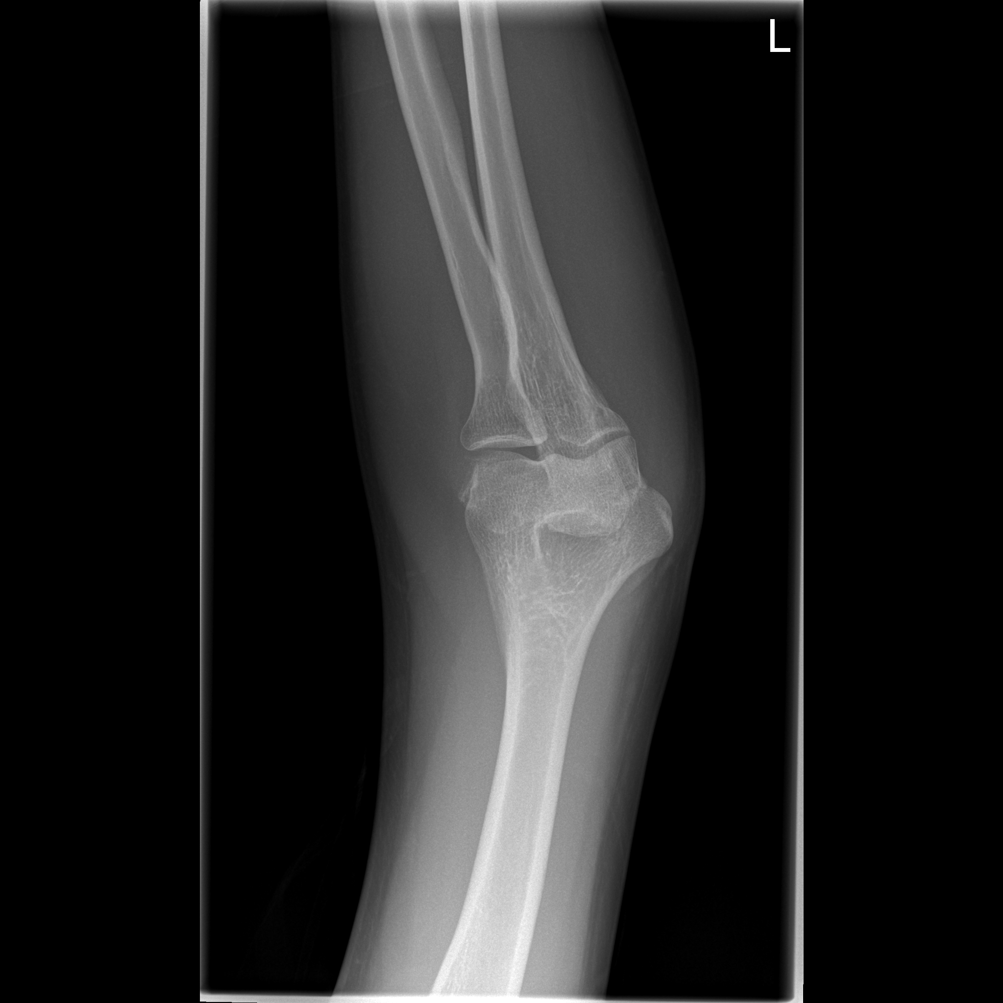
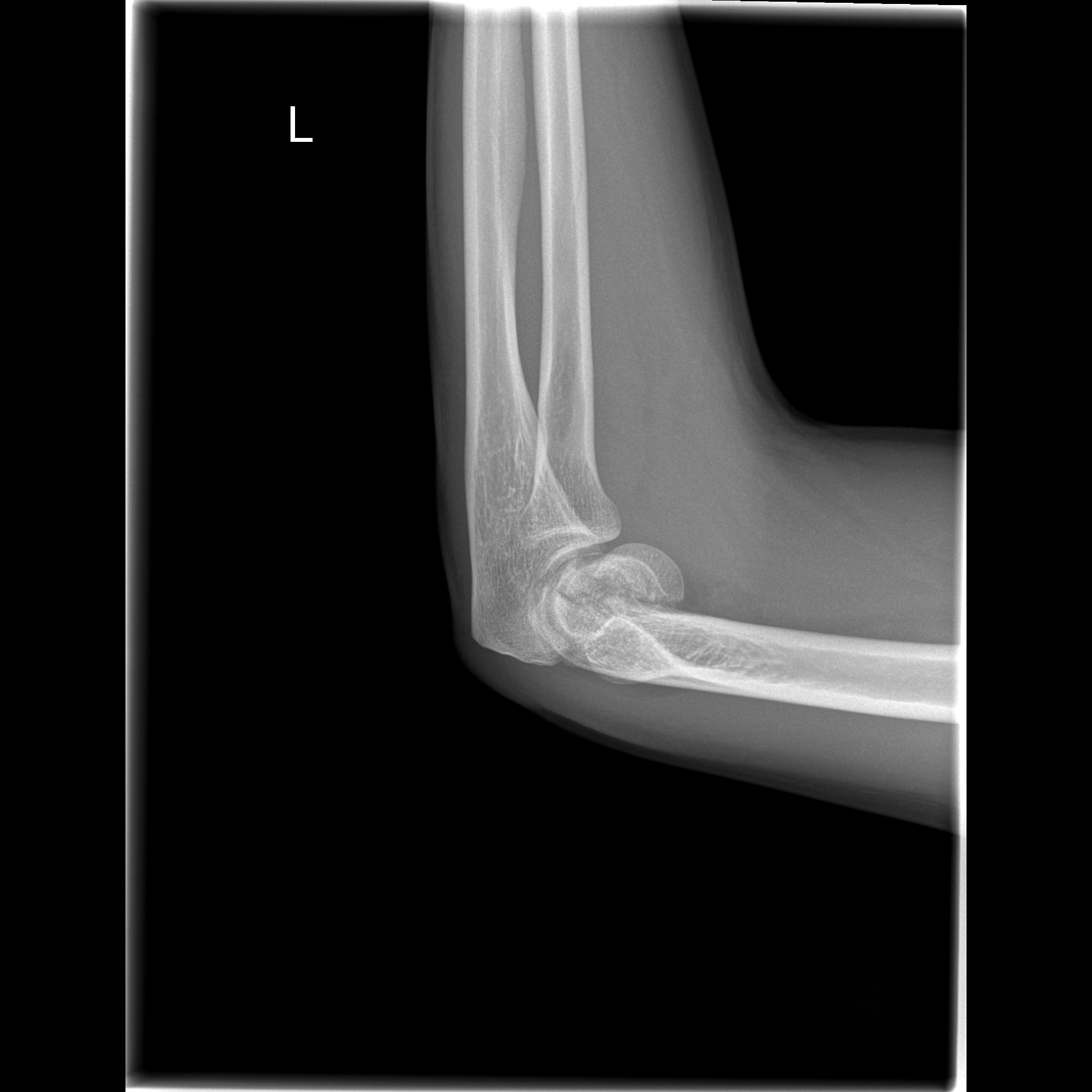
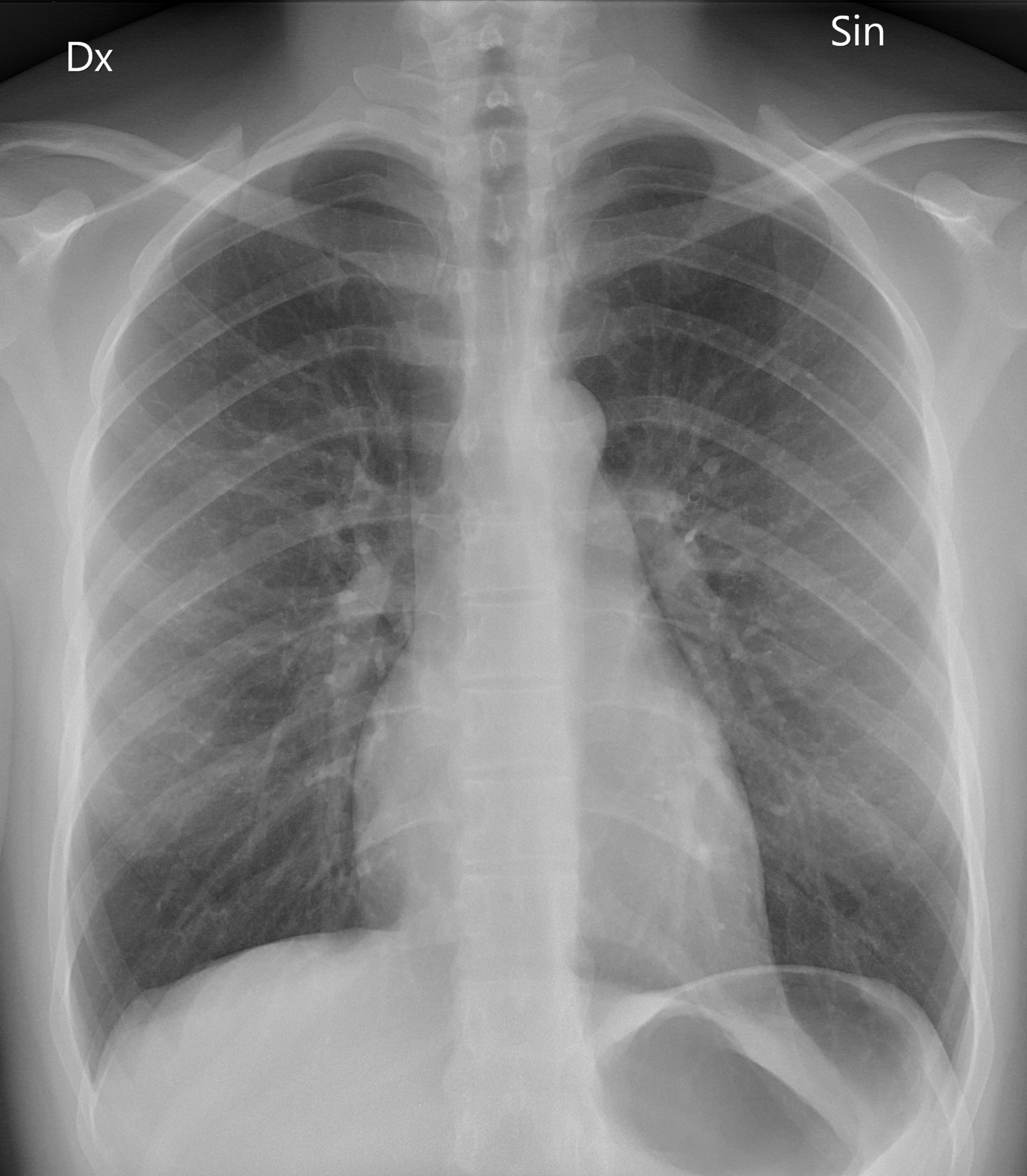
The heart is visible in an X-ray image, although it is faint without contrast.
The heart also absorbs some X-ray light, so it is possible to see it in a CT-scan. However, it does not absorb a lot of it, so the image is faint and it may be difficult to distinguish all the parts of the heart. Cardiologists are very interested to see the coronary arteries to evaluate if there are any cholesterol plaques blocking the pass of blood, but those are difficult to see in this way. To see better the coronary arteries and the heart structures, a contrasting agent can be injected in the patient’s blood. This contrasting agent absorbs the X-rays and makes the coronary arteries to be clearly visible in the image. Still, the cardiologists would like improvements on the CT image quality to better assess what they see.
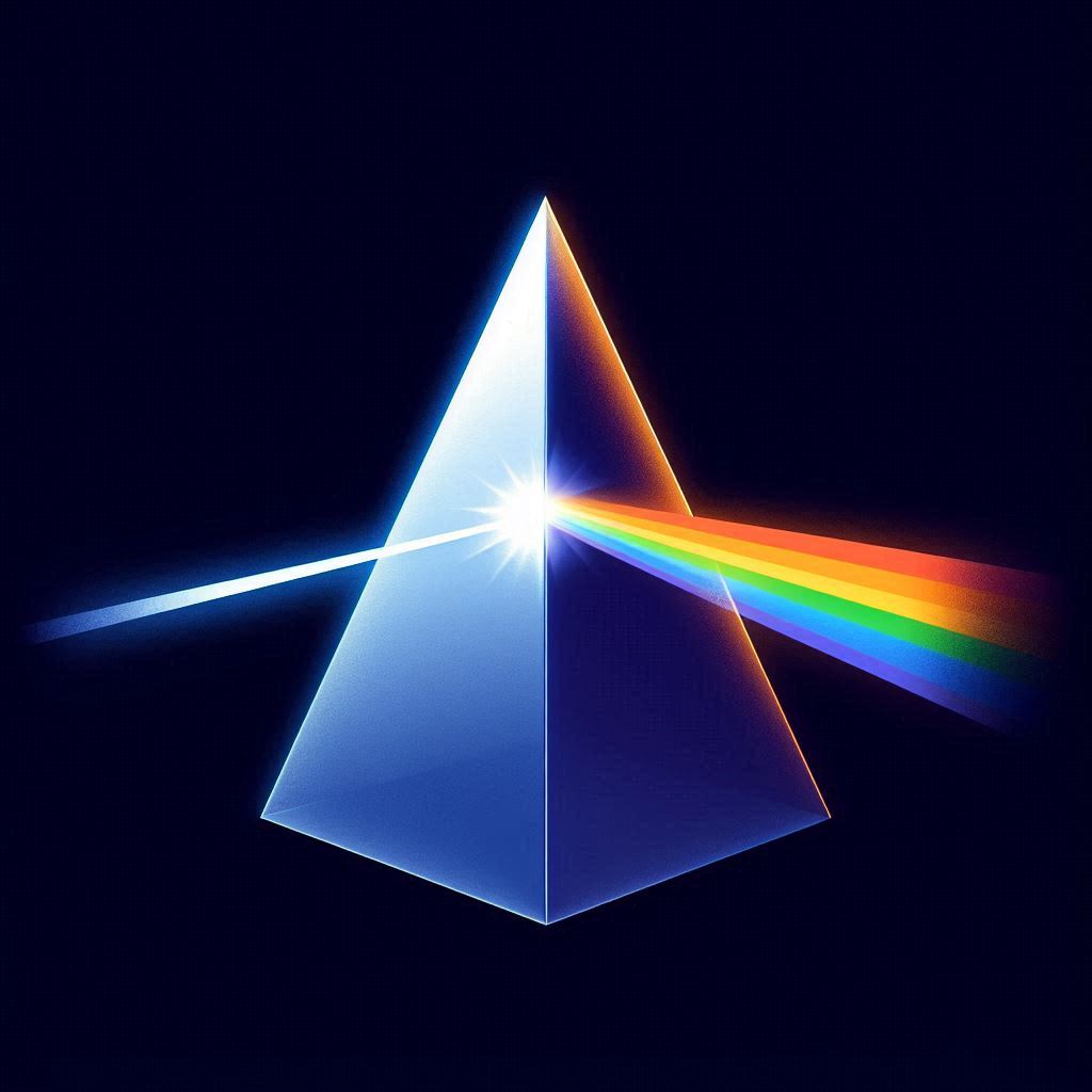
A Spectral-CT is a type of CT scan that is able to distinguish the energy of the X-rays. In the same way that visible light is composed light of different energies, from red light (which has lower energy) to violet light (which has higher energy), X-rays are also composed of higher and lower energy light. And in the same way that objects in visible light absorb certain energies and not others, which results in coloured objects, there are tissues that absorb certain energies of X-ray light better than others. In this sense, a spectral CT-scan allows us to see the ‘colour’ of X-rays, which makes CT images more informative and precise.
Status of Our Clinical Trials

The consortium has already gathered more than 10000 CT scan studies, of which 5000 are spectral CTs, and in 800 of them, the patient underwent a 2nd ischemia test. This retrospective dataset of images will be used to evaluate the performance of a diagnostic application to assess coronary injuries. With it, the sensitivity to detect coronary artery disease, as well as the specificity to rule it out, will be measured.

The recruitment of patients for the EVOLVE trial has already started. For this trial, we have identified type II diabetes patients in risk of developing coronary artery disease who could benefit from a spectral CT follow-up. AUMC, CCN and IBSAL have already started scanning patients, with more that 100 patients already included.
Our Partners
In this newsletter we will introduce two of them:

With 65 laboratories and more than 5,000 publications per year, the Université Claude Bernard Lyon 1 contributes to scientific progress in numerous fields: health, mathematics, IT, physics, chemistry, earth and space sciences, life sciences, etc. Creator of emerging knowledge and new technologies, the University is consolidating its research excellence on a global level by developing inter and multidisciplinary approaches targeting the major challenges facing society today. The leading French university in terms of the number of patents filed, Lyon 1 is an innovation factory. Ranked among the top 100 most innovative universities in the world (Reuters ranking), Lyon 1 forges solid, longterm relations with companies within the context of its proactive research transfer and partnership policy. Each year, this collaborative approach leads to the creation of new start-ups and innovative platforms as well as numerous original developments in cutting-edge fields: medicine, environment, space, etc.
UCBL, along with HCL (Hospices Civils de Lyon), leads the Work Package 2 Definitive Ischemia Diagnosis and the clinical study SPCCT-STENT, as well as participates in the studies CODEX-1 and EVOLVE.
With 200 years of medical history behind them, the Hospices Civils de Lyon are 13 public hospitals of excellence, combining all medical and surgical disciplines and driven by a threefold mission: care, research, and teaching. Together, we form a community of 23,000 women and men, both healthcare professionals and non-caregivers, sharing a single vocation: to care for and nurture each patient, regardless of their situation or illness, throughout their lives. With the purpose and strength of the commitment of our teams, united around the shared values of public service, we align our actions with a philosophy of continuous and universal care. At the heart of the city and in collaboration with all healthcare stakeholders in the Lyon region, we are a major player in the service of everyone’s health. From the care and treatment of diseases (from the most benign to the rarest), we place research at the heart of our approach to respond to today’s medical advances and anticipate tomorrow’s therapeutic challenges, thus radiating locally, nationally and internationally. Today, the 2nd largest university hospital in France, we support and train tomorrow’s medical and non-medical staff through 11 schools and institutes. Driven by a proven culture of agility that allows us to pursue our transformation efforts day after day, we are constantly innovating to team up with our patients, adopting a partner stance for and with them.
HCL, along UCBL (Université Claude Bernard Lyon 1), leads the Work Package 2 Definitive Ischemia Diagnosis and the clinical study SPCCT-STENT, as well as participates in the studies CODEX-1 and EVOLVE.
The Consortium